The clinical narrative surrounding Platelet-Rich Plasma (PRP) in orthopedics is one of captivating biological potential constantly grappling with the constraints of real-world application. PRP is not a new discovery; its principles have been known for decades, but its transition from a niche surgical adjunct to a widely marketed, non-operative treatment for musculoskeletal ailments has accelerated rapidly, often outpacing the rigorous, conclusive evidence typically demanded by evidence-based medicine. At its core, PRP is an autologous blood product, a concentrated volume of plasma derived from the patient’s own peripheral blood, characterized by a platelet count significantly exceeding baseline values. The therapeutic rationale rests on the fact that when platelets are activated at the site of injury, they release a plethora of bioactive molecules—including numerous growth factors, cytokines, and chemokines—which are instrumental in initiating and regulating the intricate processes of tissue repair, angiogenesis, and cell migration. This inherent biological safety and the appeal of utilizing the body’s own restorative agents are major drivers of its clinical popularity.
The therapeutic rationale rests on the fact that when platelets are activated at the site of injury, they release a plethora of bioactive molecules.
The fundamental challenge in evaluating PRP’s true efficacy lies in its profound heterogeneity; it is not a standardized drug but a variable biological commodity. The term ‘PRP’ represents a diverse family of products, each differing substantially based on the preparation protocol employed. These variations stem from factors such as the initial volume of blood drawn, the specifics of the centrifugation protocol (speed, time, number of cycles), and the use of proprietary commercial devices versus custom lab preparations. Consequently, the resulting product can differ dramatically in its final platelet concentration, the presence or absence of leukocytes (white blood cells), and the volume of the injectable solution. This lack of a unified formulation means that a positive result reported in one study utilizing a Leukocyte-Rich PRP (LR-PRP) for a chronic tendinopathy is not easily transferable to a clinic using a Leukocyte-Poor PRP (LP-PRP) for intra-articular osteoarthritis. These methodological inconsistencies are precisely why the current body of literature contains so many conflicting outcomes, frustrating attempts at definitive meta-analyses and universally accepted treatment guidelines.
The fundamental challenge in evaluating PRP’s true efficacy lies in its profound heterogeneity; it is not a standardized drug but a variable biological commodity.
To bring some order to this chaos, classification systems have been proposed by various researchers, attempting to categorize PRP based on its essential quantifiable characteristics. Systems like the DEPA classification (Dose of injected platelets, Efficiency of production, Purity, and Activation) or the PAW classification (Platelets, Activation, and White cells) seek to provide a common language for reporting and comparing studies. These models acknowledge that the sheer concentration of platelets alone is insufficient; the final dose delivered to the target tissue, the concentration of accompanying white blood cells, and the mechanism by which the product is activated—or not activated—all fundamentally dictate the final biological signal imparted to the injury site. A higher absolute dose of platelets is generally considered necessary to achieve a biological effect that significantly surpasses the body’s natural healing response. The orthopedic physician must look beyond the generic label of ‘PRP’ and consider which specific formulation is most appropriate for a given tissue pathology and healing goal.
These models acknowledge that the sheer concentration of platelets alone is insufficient; the final dose delivered to the target tissue, the concentration of accompanying white blood cells, and the mechanism by which the product is activated—or not activated—all fundamentally dictate the final biological signal imparted to the injury site.
The role of leukocytes in the PRP formulation is a particularly contentious subject, representing a crucial biological trade-off. Leukocyte-Rich PRP (LR-PRP), while containing a higher concentration of all cellular components, also carries a load of neutrophils and macrophages. These cells are essential in the initial inflammatory phase of healing and contain lytic enzymes and antimicrobial peptides. For certain chronic tendon pathologies, which are often characterized by a stalled or incomplete inflammatory response, the pro-inflammatory stimulus from LR-PRP may be desirable to “re-boot” the healing cascade. However, in the delicate environment of a synovial joint affected by osteoarthritis, the introduction of excess leukocytes may be counterproductive. Leukocytes can release catabolic enzymes and pro-inflammatory cytokines such as Interleukin-1$\beta$ and Tumor Necrosis Factor-, which can potentially accelerate cartilage degradation and increase post-injection pain and swelling. Therefore, many clinicians favor Leukocyte-Poor PRP (LP-PRP) for intra-articular injections to focus purely on the regenerative and anti-inflammatory properties of the concentrated platelets without the undesirable catabolic load.
For certain chronic tendon pathologies, which are often characterized by a stalled or incomplete inflammatory response, the pro-inflammatory stimulus from LR-PRP may be desirable to “re-boot” the healing cascade.
In the realm of chronic tendinopathies, such as gluteal tendinopathy or chronic patellar tendonitis, the clinical data suggests that PRP, when properly characterized and applied, offers a sustained advantage over traditional therapies. These injuries are often avascular, experiencing limited natural healing. The concentrated delivery of growth factors like TGF- and PDGF acts as a biochemical scaffold and signaling hub, promoting the local proliferation of tenocytes and enhancing the synthesis and structural organization of new collagen fibers, ultimately attempting to restore the viscoelastic properties of the tendon. The comparative studies often demonstrate that while corticosteroid injections may provide superior short-term pain relief, they frequently lead to relapse and may be associated with long-term tissue weakening. PRP, conversely, tends to show a slower initial response but a more durable and functionally significant improvement at the six-month to one-year follow-up, suggesting a true restorative effect rather than mere symptomatic masking.
PRP, conversely, tends to show a slower initial response but a more durable and functionally significant improvement at the six-month to one-year follow-up.
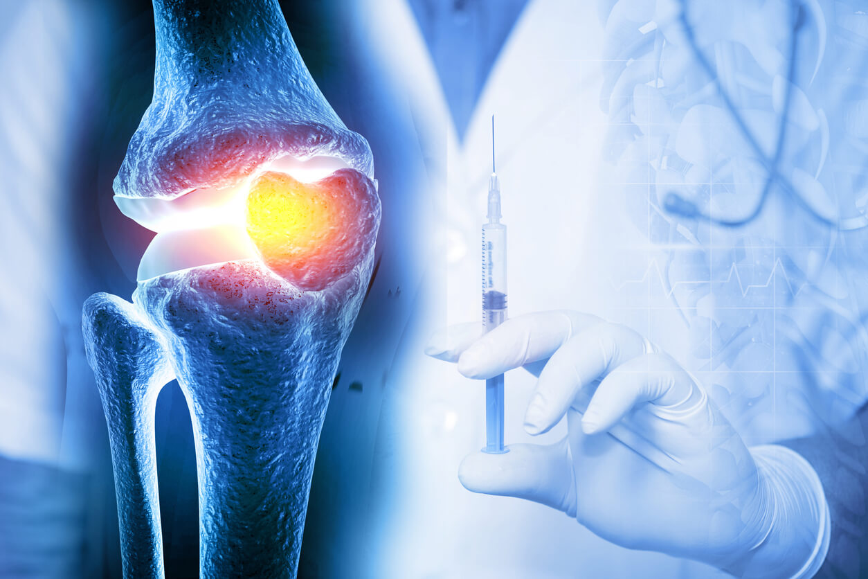
For knee osteoarthritis (OA), the evidence base has matured significantly, positioning intra-articular PRP as a viable non-surgical option for mild to moderate cases, particularly in younger, active patients aiming for joint preservation. The treatment acts not only by stimulating the anabolic activity of chondrocytes but also through a critical immunomodulatory effect on the synovial fluid. PRP components can help to quench the chronic, low-grade inflammatory state within the joint, a primary driver of pain and further cartilage breakdown. While PRP does not regenerate lost cartilage structure in a radiographically verifiable sense, multiple high-level trials have reported substantial and clinically significant improvements in pain scores and functional outcomes, often surpassing the effects observed with hyaluronic acid injections or saline placebos over equivalent time frames. The duration of this symptomatic relief, however, remains variable and often necessitates repeat injections to maintain the benefit.
The treatment acts not only by stimulating the anabolic activity of chondrocytes but also through a critical immunomodulatory effect on the synovial fluid.
The integration of PRP into the surgical setting, primarily as a biological augment to enhance healing at bone-tendon or bone-ligament interfaces, offers another promising avenue. In procedures like rotator cuff repair, where the risk of tendon re-tear is significant, or in ligament reconstructions, the local application of PRP is designed to accelerate the biological fixation of the graft or repaired tissue. By providing a high local concentration of factors that promote tissue ingrowth and vascularization, PRP theoretically strengthens the weakest link in the repair construct. While the mechanical benefits on a population level in large-scale surgical trials have been inconsistent, the underlying biological mechanism remains sound, leading many surgeons to selectively employ PRP, particularly in high-risk patients or when dealing with larger tears where the biological demand for healing is highest. This application highlights the product’s utility not as a standalone treatment but as a supportive biological tool.
By providing a high local concentration of factors that promote tissue ingrowth and vascularization, PRP theoretically strengthens the weakest link in the repair construct.
The patient-specific factors are arguably as crucial as the product composition itself, introducing another layer of inherent complexity. The efficacy of an autologous therapy like PRP is fundamentally dependent on the biological quality of the patient’s own blood. Variables such as age, nutritional status, systemic disease (e.g., diabetes), and the use of certain medications (e.g., NSAIDs, anticoagulants) can all influence the functionality and growth factor content of the harvested platelets. A patient with a significant underlying inflammatory condition may yield a PRP product that is inherently less potent or contains a disproportionate amount of inflammatory cells, thereby potentially limiting the therapeutic outcome irrespective of the precision of the preparation technique. This dependency underscores the need for a comprehensive pre-treatment patient assessment and counseling, moving the approach toward a model of personalized orthobiologics.
The efficacy of an autologous therapy like PRP is fundamentally dependent on the biological quality of the patient’s own blood.
Looking forward, the future of PRP in orthopedics is tied to the evolution of precision medicine. The current clinical uncertainty necessitates a shift towards rigorous phenotyping of both the patient and the PRP product. Advanced research is focusing on developing assays that can rapidly characterize the functional capacity of the harvested platelets and their secreted growth factor profile. Furthermore, incorporating artificial intelligence and machine learning to correlate specific PRP compositions (e.g., absolute platelet dose, leukocyte ratio) with long-term clinical outcomes for distinct musculoskeletal pathologies is essential. This data-driven approach aims to move beyond generic guidelines, allowing clinicians to tailor the PRP formulation—or choose a different orthobiologic entirely—to the unique biological requirements of the individual patient and their specific injury, thereby maximizing the probability of a successful therapeutic response.
This data-driven approach aims to move beyond generic guidelines, allowing clinicians to tailor the PRP formulation—or choose a different orthobiologic entirely.
The journey of PRP from an experimental concept to a mainstream orthopedic treatment is a testament to the power of endogenous healing, yet it remains a work in progress. For it to solidify its position as an indispensable, first-line treatment, the medical community must overcome the persistent barriers of standardization and methodological ambiguity. The final determination of PRP’s role will not rest on a single, universal finding, but on the capacity to precisely match a characterized biological product to a defined pathological state, supported by long-term, functional evidence that clearly delineates its superiority over existing conservative options.
